


Sometimes you might encounter a small lump or bump on your gum which might be painful. It might sound alarming, but it does not always mean something serious and concerning. So does it mean all lumps on gum should be ignored? Well the answer is no! Gum bumps range from harmless, self-resolving sores or urgent dental infections requiring same-day treatment or sometimes. At times longstanding, progressive bumps on the gum might be hinting at a more serious underlying problem like oral cancer spreading silently within the jaw. Understanding what type of bump you have on the gum is the first step toward knowing what to do next.
This guide covers common causes of gum bumps, evidence-based treatment options, a practical self-examination guide, and prevention strategies that most online resources overlook entirely.
What Is a Bump on the Gums?
A bump or lump on the gums is any raised area of tissue along the gum line. It may be soft or hard, painful or painless, white, red, or flesh-colored with size ranging from a few millimetres to several centimeters. These factors become essential as they help your dentist determine and narrow down upon what might be causing the problem. Amongst this the key clinical indicator characteristics of a bump include its size, color, texture, location, and how long it has been present.
Some bumps reflect purely local problems (an injury, a trapped food particle), while others are the visible surface sign of a deeper infection or, rarely, a cancerous process.
Why do I have a bump on my Gums?
Common causes of bump on the gums might range from something as simple as infections to complicated conditions such as oral cancer. Let's discuss the causes which might lead to bump on the gums.
1. Dental Abscess / Gum Abscess
A gum abscess is a small and localized collection of pus in the gum region. It is caused by a bacterial infection. Now, this bacterial infection might be originating from the tooth itself followed by its spread to the gums. Or it might be directly originating from the infection within gums. Broadly it can be classified as
- Periapical abscess - originates at the tip of a tooth's root, usually from untreated tooth decay or a cracked tooth. Following which the infection spreads into the supporting structures around the tooth.
- Gingival abscess - forms in the gum tissue itself. Tooth is not the source of infection. It typically is a complication of advanced gum disease.
Symptoms
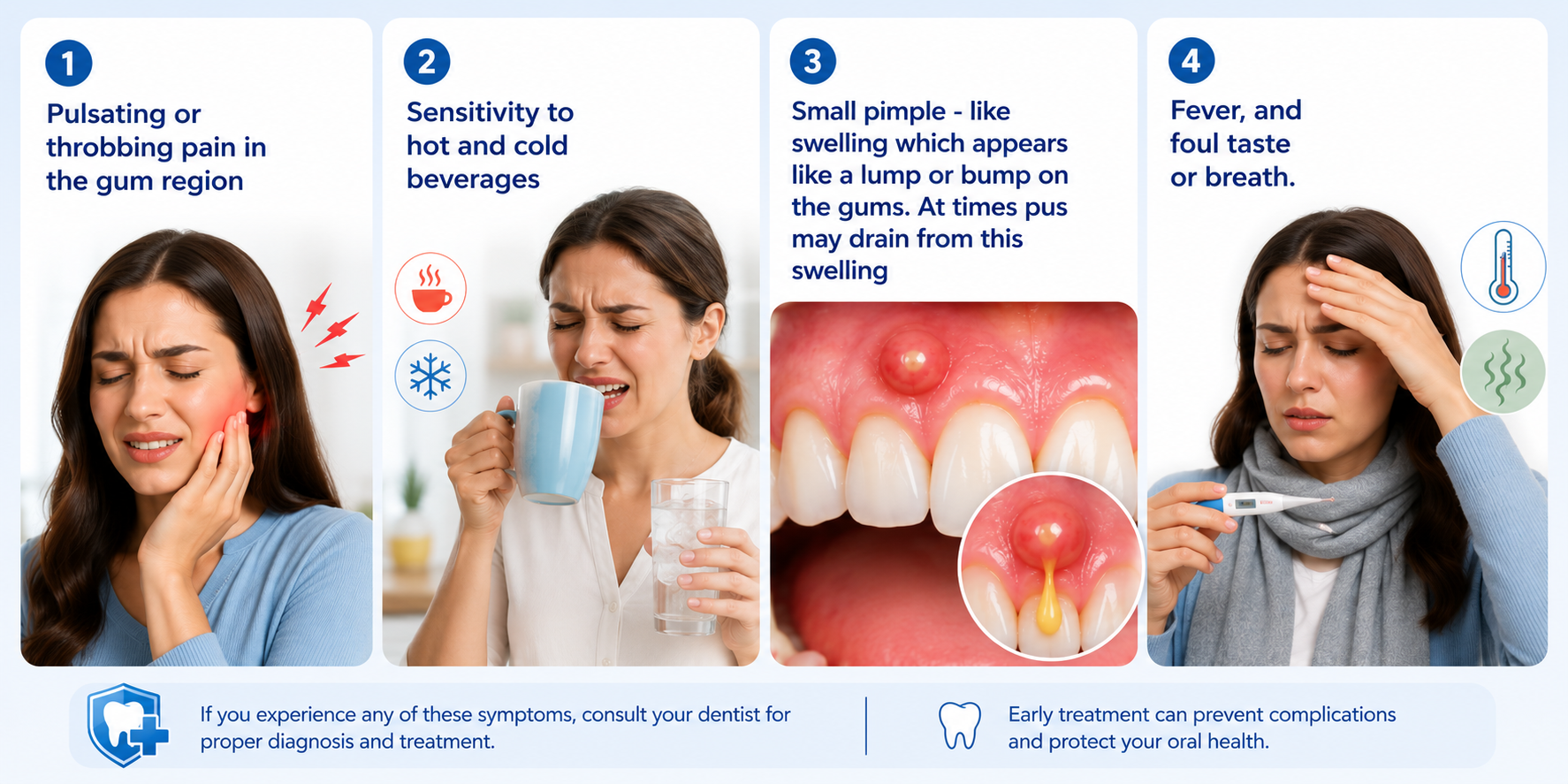
Treatment
Antibiotics, drainage, root canal therapy, or, in severe cases, tooth extraction.
2. Canker Sores (Aphthous Ulcers)
Canker sores are among the most common oral lesions, affecting approximately 20% of the general population. They appear as small, round ulcers with a white or yellow center surrounded by a red border. They are extremely painful and might vary in their numbers ranging from one to several.
They are not contagious and can be triggered by several factors like :
- Minor mouth injuries (dental treatment, sharp food injury, biting the cheek)
- Emotional stress
- Nutritional deficiencies (vitamin B12, iron, folate)
- Certain foods (citrus, nuts, chocolate)
Treatment
They typically resolve on their own within 7–14 days. No treatment can permanently prevent recurrence as they might be triggered due to several reasons. However a few things might be recommended by your dentist to reduce pain and provide relief from discomfort. These include-
- Topical anesthetics
- antimicrobial rinses
- corticosteroid gels
3. Dental Cysts
Dental cysts can be understood as fluid-filled sacs that develop in or around the jaws and gums. They grow slowly, are often painless, and may go unnoticed for years until detected on a dental X-ray by chance or while dental treatments.
They are most commonly associated with an unerupted tooth. Common types of dental cysts include:
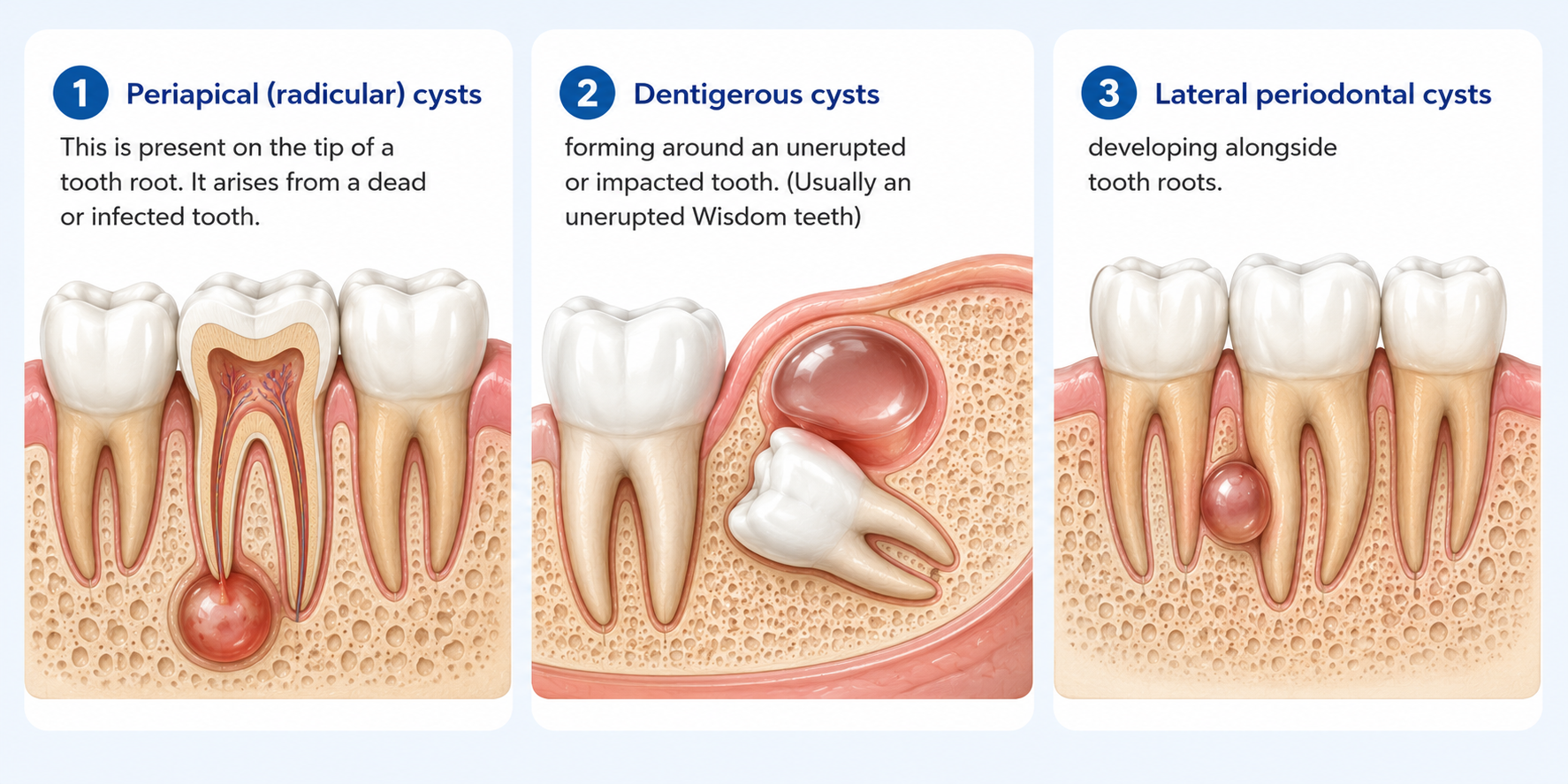
It is important to remember that these cysts are often painless and thus ignored. At times they are detected at a very late stage when their size has increased to several centimetres. Thus this emphasises the fact that routine dental visits every 6 months are very essential to detect silent swelling of the jaws timely. If left untreated, dental cysts can expand to displace teeth and resorb jawbone or might become infected leading to severe pain.
Treatment
Involves surgical removal (enucleation) or marsupialization for larger cysts in which your oral surgeon creates a small opening in the cyst and sutures the edges to keep it open, allowing the fluid to drain gradually thereby reducing the size of the cavity.
4. Irritational Fibroma
A gingival fibroma is an overgrowth of fibrous tissue on the gum region which arises in response to a long standing irritation or trauma. This irritation may be arising from a sharp tooth or a dental filling which is overhanging and hitting against the gum ill fitting dentures, braces. It is one of the most common benign soft-tissue lesions in the oral cavity . You might seen them having the following appearance :
- Hard, smooth, dome-shaped lumps on the gums
- Either flesh like in colour or slightly whiter appearance than surrounding gum
- Painless and slow-growing lumps
Treatment
Fibromas do not resolve on their own. Surgical removal by your dentist is the definitive treatment, and the irritating cause which is repeatedly cursing trauma (ill-fitting appliance) should also be identified and corrected to prevent recurrence.
5. Pyogenic Granuloma
Pyogenic granuloma is a harmless overgrowth of gum tissue that bleeds easily (even on slightest touch) and can grow rapidly in a short duration attatining lagre sizes. . The exact cause is unclear, but researchers link it to trauma, hormonal changes, infections, and certain medications . One of the subtypes of pyogenic granuloma known as "pregnancy tumor" (epulis gravidarum), occurs in up to 5% of pregnant women due to hormonal variations seen in pregnancy. These elevated and altered levels of hormones exaggerate the gum tissue's response to local irritants.
Despite its alarming name, it is entirely non-cancerous.
Treatment
Lesions often regress after delivery, but persistent ones require surgical removal by an experienced dentist. Recent advances use Laser to remove the growth which allso speeds up the rate of recovery.
6. Mandibular Torus (Oral Tori)
A mandibular torus is a slow-growing, non cancerous bony prominence that develops on the inner surface of the lower jaw, covered by normal gum tissue. Tori are frequently bilateral. The key causative factors include genetics, teeth grinding, and dietary calcium levels.
Treatment
Most tori require no treatment. Removal is indicated only when the torus interferes with speech, swallowing, denture fitting, or oral hygiene maintenance.
7. Pericoronitis
It is the most frequently overlooked cause which appears as a bump on gum near the wisdom tooth . Pericoronitis is an inflammatory condition affecting the gum tissue surrounding a partially erupted tooth. Most often it is seen associated with a wisdom tooth which has not fully erupted in the moth. As a wisdom tooth partially erupts in the mouth it is frequently covered by a flap of gum tissue (operculum). This flap is a common site for food accumulation. Bacteria proliferate in this warm, moist environment, triggering infection. You might notice the following
- A swollen, tender flap of gum behind the last molar
- Difficulty opening the mouth (trismus)
- Pain that radiates to the ear, jaw, or throat
- Fever in severe cases
If left untreated, pericoronitis can progress to life-threatening deep-space infections, including Ludwig's angina.
Treatment
The area is professionally cleaned by your dentist. The flap of gum covering the tooth is removed. In cases where frequent recurrence is reported the dentist might need to remove the tooth as well.
8. Oral Cancer
While most gum bumps are non cancerous (Benign), a small percentage represent cancerous disease. Gum cancer is most commonly squamous cell carcinoma (SCC) of the gingival tissue. Early lesions may appear as a non-healing sore, a white or red patch, or a firm, irregular bump. Any growth persisting for a duration of more than two weeks either painless or painful needs to be examined by your Dentist.
Key risk factors include which might make you more prone to oral cancer include -
- Tobacco use
- Alcohol consumption
- Human papillomavirus (HPV) infections - particularly HPV-16 and HPV-18, linked to oropharyngeal cancers
- Age over 55
- Male sex
Early detection through regular dental screenings is the single most impactful preventive measure.
Systemic Health Conditions might be causing bump on your gums
Oral health is closely linked to your general health rather than functioning independently. As it is rightly termed that Mouth is the mirror of your body’s health. Few of the diseases affecting your body might thus be manifesting their symptoms in the oral cavity silently. Lets try and understand this using a few examples.
- Diabetes: Increased blood sugar levels cause a decrease in the immune cell function and reduces the body's ability to fight oral bacterial infections, making abscesses and periodontal disease more common and harder to treat.
- HIV/Immunosuppression: Patients on immunosuppressive therapy or living with HIV are more susceptible to opportunistic oral infections, including bacterial, viral and fungal infection.
- Pregnancy: Hormonal changes (estrogen and progesterone surges) exaggerate gum tissue responses to irritants, increasing the risk of lumps and bumps on the gingiva
If you have any of these conditions, or even if you are feeling healthy but you notice a new gum bump on your gums, which is increasing progressively in size or painfulconsult a healthcare provider immediately. Sometimes what might appear as small and insignificant might require immediate attention before things get complicated.
How to Examine Your Gums at Home?
Regular self-examination takes less than two minutes and can help you detect any changes in the mouth early. Faster is the disease identified better are the disease outcomes.The following method is adapted from the Mouth Cancer Foundation's recommended protocol:
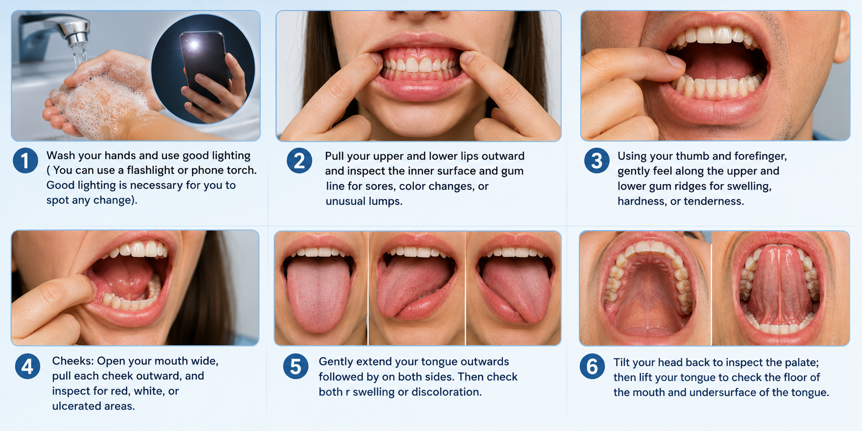
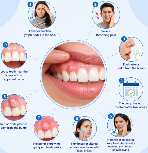
See a dentist promptly if you notice:
- If the bump is lasting for more than two weeks or progressively increasing in its size.
- Unexplained bleeding from the bump/ lump on the gum
- Numbness or tingling in the gums or lips
- Difficulty swallowing or opening your mouth
- Fever
How to get rid of Bump on gums?
At-Home Management (for mild, non-infectious causes)
When a bump is likely a minor canker sore or irritation, the following measures can provide relief:
- Warm saltwater rinses (half a teaspoon of salt in 8 oz of lukewarm water). It reduces inflammation and bacterial load thereby promoting healing.
- Soft-bristle toothbrush
- Which will avoid further trauma
- Avoid spicy, acidic, or hard foods that may irritate the area
Professional Treatments
| Condition | Treatment |
|---|---|
| Gum Abscess | Antibiotics + drainage + root canal or extraction |
| Canker Sore/Ulcers | Topical corticosteroids, B12/folate supplementation |
| Dental Cyst | Surgical enucleation or marsupialization |
| Fibroma | Surgical excision |
| Pyogenic Granuloma | Surgical excision; watchful waiting in pregnancy |
| Mandibular Torus | Surgical removal if functionally impaired |
| Pericoronitis | Irrigation, antibiotics, possible extraction |
| Oral Cancer | Surgery, radiation, chemotherapy (often combined) |
How to prevent gum bumps from occurring?
Prevention is the most effective and the most under-discussed aspect of gum bump management. Minor changes in your oral health and hygiene maintenance might prevent the occurrence of these bumps.
- Brush twice daily and floss once daily to prevent plaque accumulation that feeds bacterial infections
- Attend dental check-ups every six months. This will help in the early detection of lesions, cysts, and cancers dramatically improves outcomes.
- Quit tobacco in all forms : cessation significantly reduces oral cancer risk even after years of use
- Abstain from alcohol consumption : combined tobacco and alcohol use produces a synergistic increase in oral cancer risk
- HPV vaccination : although developed for cervical cancer, HPV vaccines offer broader protection including against HPV- associated oral and oropharyngeal cancers.
- Ensure adequate nutrition : maintain sufficient vitamin B12, iron, and folate levels to reduce canker sore frequency
- Keep dental appliances well-fitted : ill-fitting dentures and braces are leading irritants that cause fibromas
- Wear a night guard if you grind your teeth (bruxism), reducing torus formation risk
When to See a Dentist for a bump on the gums?
Seek dental care as soon as possible if you experience any of the following alongside a gum bump:
It is important to understand that sometimes the bump might sometimes drain pus or blood and shrink in size. However, unlike skin pimples, this does not mean that the infection is drained and over. Dental infection can only be cured by management of the underlying cause or else the bump will reoccur.
Conclusion
A bump or lump on the gum is a common occurrence. At times you may notice a pain accompanied with the bump and at times it might be completely silent, slowly progressing leading to difficulty in opening mouth or swallowing. Reasons can range from minor infections to something more serious like oral cancer. What is important to understand here is that you should not be ignoring it. If you notice any such bump on your Gum, immediately visit your dentist for appropriate treatment. Blind self attempts of managing it yourself or delaying the treatment might prove more harmful than you can imagine.

Not every oral bump appears on the gums learn what those bumps on the back of your tongue could be trying to tell you.
Frequently Asked Questions
Can a bump on my gum go away on its own?
It depends on the cause. Canker sores and minor pyogenic granulomas can resolve without treatment. Abscesses, cysts, and fibromas will not, and oral cancer requires prompt intervention.
Is every gum bump an infection?
No. Many gum bumps ( including fibromas, mandibular tori, and canker sores) are not infections at all. However, gum abscesses and pericoronitis are bacterial infections and require antibiotics or surgical management.
Should I pop a gum bump?
Never. Attempting to pop a gum bump can spread bacteria deeper into surrounding tissue, potentially escalating a localized infection into a serious systemic problem.
What does an oral cancer bump look like?
Oral cancer bumps often appear as firm, irregular lumps with poorly defined borders. They may be red (erythroplakia), white (leukoplakia), or mixed. They typically do not hurt in early stages which is why regular dental screening is critical.
Scientific References
- Chavan, M., et al. (2012). Recurrent aphthous stomatitis: A review. Journal of Oral Pathology & Medicine, 41(8), 577–583.
- Coletta, R. D., Yeudall, W. A., & Salo, T. (2024). Current trends on prevalence, risk factors and prevention of oral cancer. Frontiers in Oral Health, 5, 1505833.
- Dangore-Khasbage, S., & Teredesai, T. (2026). Association between mandibular third molar position and recurrent pericoronitis: Protocol for a cross-sectional study. JMIR Research Protocols.
- Fatani, et al. (2024). Irritational fibroma mimicking an odontogenic infection: A case report of a misdiagnosed extraoral fibroma. Cureus, 16(3), e56311.
- Herrera, D., et al. (2018). Acute periodontal lesions (periodontal abscesses and necrotizing periodontal diseases) and endo-periodontal lesions. Journal of Clinical Periodontology, 45(Suppl 20), S78–S94.
- Igoumenakis, D., et al. (2022). Pericoronitis. In StatPearls. National Center for Biotechnology Information.
- Jafarzadeh, H., Sanatkhani, M., & Mohtasham, N. (2006). Oral pyogenic granuloma: A review. Journal of Oral Science, 48(4), 167–175.
- Neville, B. W., Damm, D. D., Allen, C. M., & Chi, A. C. (2015). Oral and Maxillofacial Pathology (4th ed.). Elsevier Saunders.
- Oral Tori Pilot Study. (2024). Oral tori findings in an adult Albanian population: A single-center pilot study. PMC.
- Rich, B. J., Samuels, S. E., Azzam, G. A., Kubicek, G., & Freedman, L. (2024). Oral cavity squamous cell carcinoma: Review of pathology, diagnosis, and management. Critical Reviews in Oncogenesis, 29(3), 5–24.


Member discussion